Artificial Intelligence (AI) integrated within a Portable 3D ABUS Device
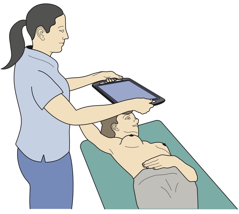
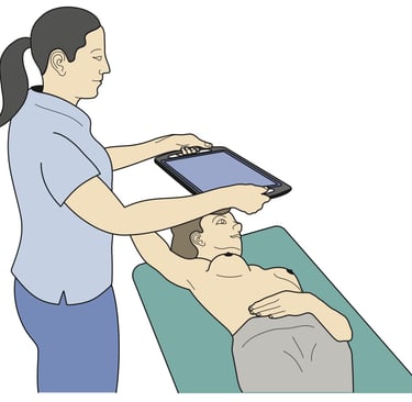
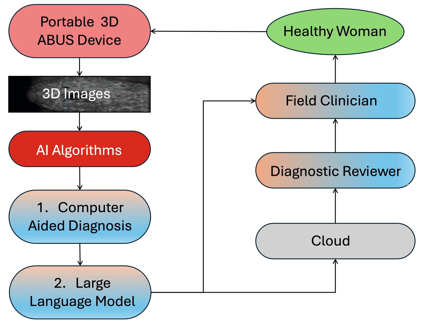
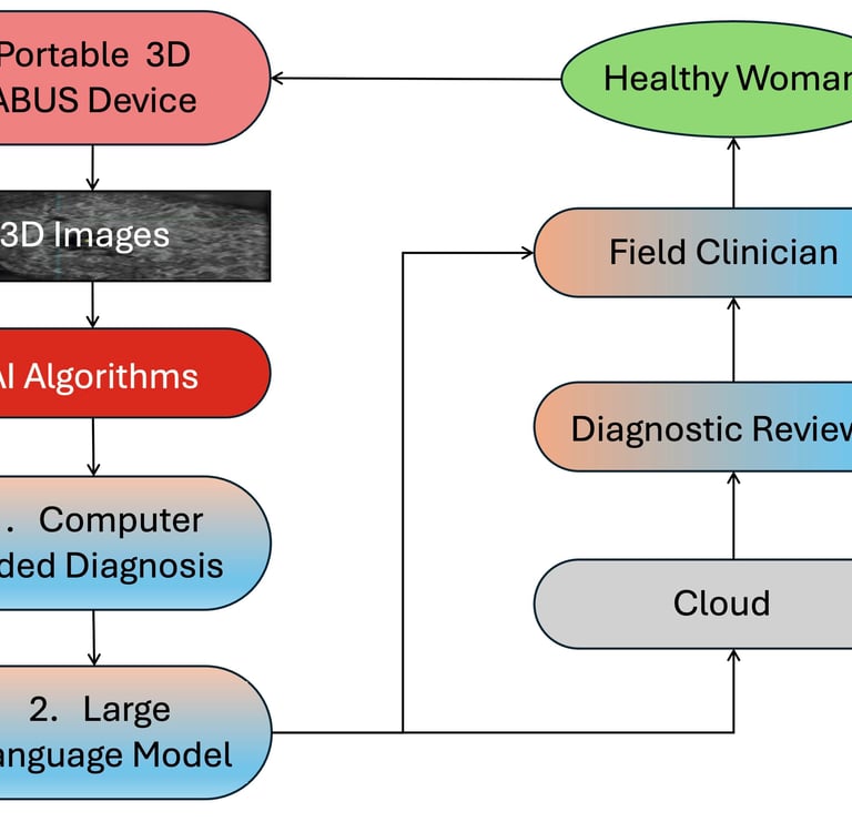
The first diagram shows a field clinician moving the portable 3D ABUS device over the woman's breast, while the adjacent flow diagram demonstrates how the 3D system will be applied in the field. The clinician engages with a healthy woman volunteer using the portable device which generates 3D images of each breast. There are two places in the data flow where AI plays an important role after acquisition of the 3D images and artefacts have been removed: (1) an algorithm based on convolutional neural network (CNN) technology is applied to detect breast lesions; and (2) a large language model (LLM) interacts with both the diagnostic reviewer (i.e. radiologist) and the field clinician. If a malignant lesion is detected, the woman will be referred for further testing and treatment.
Innovative Cancer Detection
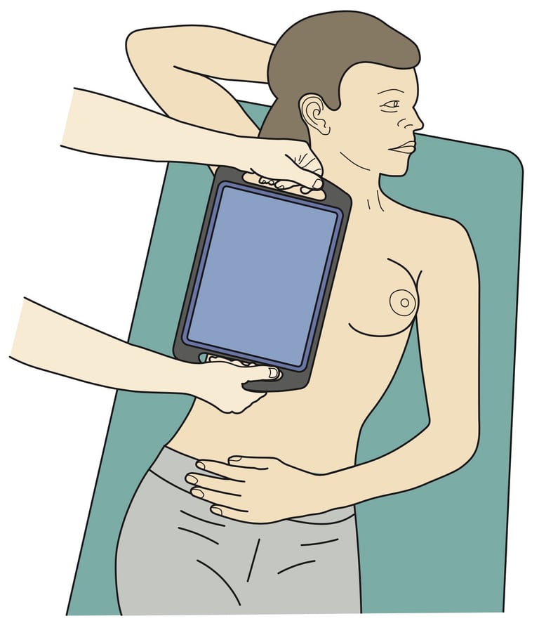
The field clinician places the 3D ABUS device on the woman's breast. At this stage the screen on the tablet computer is blank.

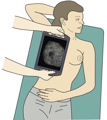
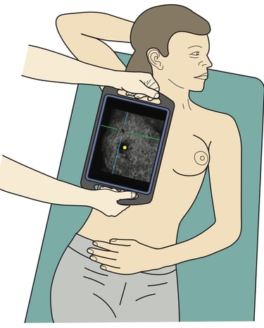
The clinician instructs the device to acquire an image of the breast tissue and immediately a coronal plane view appears on the screen.
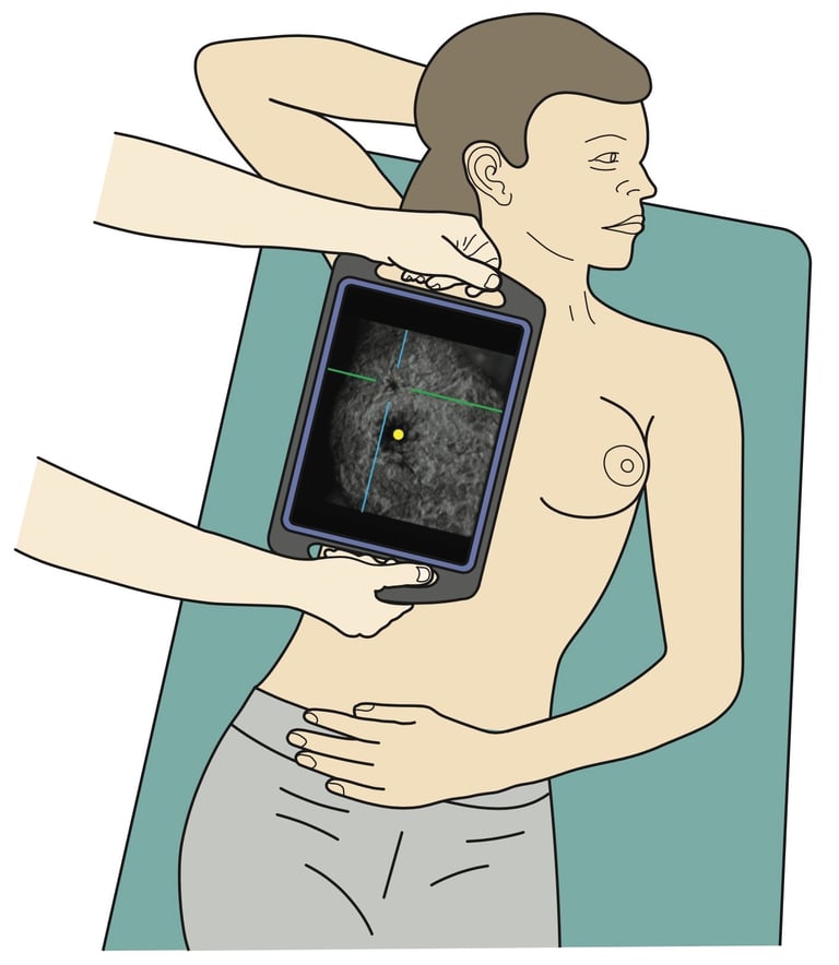
Next she turns on the AI algorithm which highlights the nipple (yellow dot) and identifies the malignant lesion in the cross hairs.
Innovation
Revolutionising breast cancer detection with AI technology.
© 2026. All rights reserved.